
Typically sleep apnea events last for 30–60 s. Obstructive sleep apnea is a sleep disorder characterized by repetitive cessations of respiratory flow for at least 10 s. The evaluation of sleep is still based on visual inspection of all traces since automatic analysis of sleep recordings still has many limitations not acceptable to the sleep physician ( Penzel and Conradt, 2000). Today's sleep laboratories use computer-based equipment that allows digital recording, evaluation, and archiving of the sleep records. The electrocardiogram (ECG) is recorded routinely, EMG of both legs is recorded to detect movement disorders during sleep. Respiration is recorded with respiratory effort sensors at the chest and the abdomen, effective oronasal airflow with thermistor sensors or as recently discussed with nasal pressure transducers ( Hosselet et al, 1998), snoring with a laryngeal microphone and oxygen saturation derived from a finger or ear oximetry. Today sleep laboratories perform cardiorespiratory sleep studies ( ATS, 1989), which adds the recording of respiration and further signals.

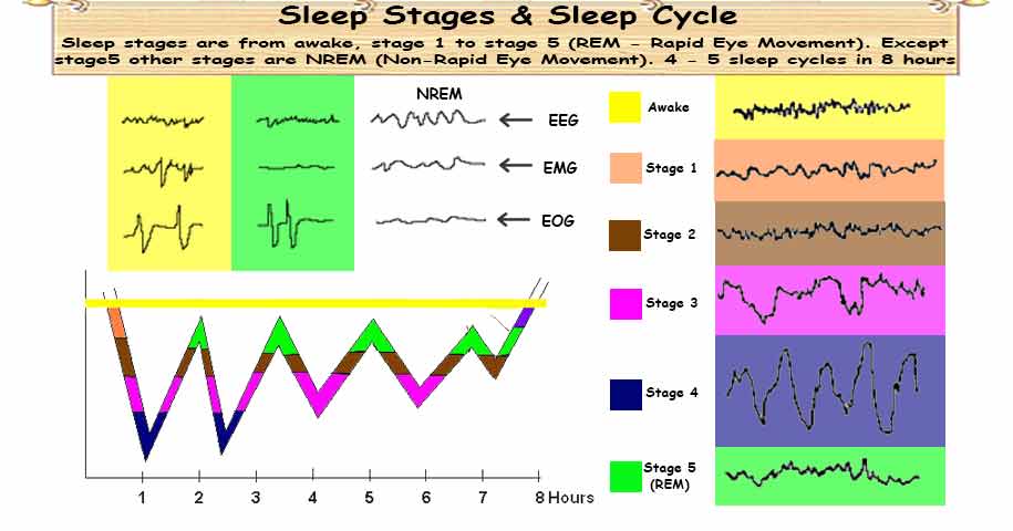
A normal night consists of six sleep cycles where the proportion of deep sleep decreases from the beginning to the end of the night and the proportion of REM sleep increases at the same time. Such a sequence is called a sleep cycle which has a typical duration of 90–110 min. In normal sleep, the stages follow a structured sequence starting with wake, then light sleep with stages 1 and 2, followed by deep sleep (slow wave sleep) with stages 3 and 4, and then followed by REM sleep. Each 30-s epoch of the sleep recording is labeled with one sleep stage. These sleep stages are evaluated in consecutive 30-s epochs. Characteristic for REM sleep are the REMs. During ‘rapid-eye-movement (REM) sleep’ EMG amplitude drops to its lowest values and the EEG is wake like with possible sawtooth waves but no alpha waves. Sleep stages were defined as ‘wake’ with dominant beta and alpha waves, ‘stage 1’ with less alpha waves slow rolling eye movements, ‘stage 2’ with sleep spindles, K-complexes, some theta waves, some delta waves, ‘stage 3’ with up to 50% delta waves, ‘stage 4’ with more than 50% delta waves, and low EMG amplitude. The technique of sleep recording and the definition of sleep stages has been given in recommendations compiled by Rechtschaffen and Kales (1968) and these are still regarded as the universal common language of sleep medicine. The internal structure can be described by sleep stages that are differentiated based on typical patterns and waves found in the electroencephalography (EEG), electro-oculography (EOG), and electromyography (EMG) recordings. Sleep is not just the absence of wakefulness but has an own internal structure.


 0 kommentar(er)
